Research Lab Results
-
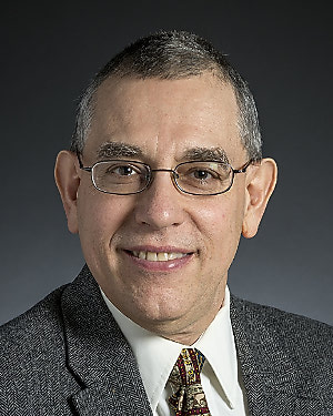
J. Marie Hardwick Laboratory
Our research is focused on understanding the basic mechanisms of programmed cell death in disease pathogenesis. Billions of cells die per day in the human body. Like cell division and differentiation, cell death is also critical for normal development and maintenance of healthy tissues. Apoptosis and other forms of cell death are required for trimming excess, expired and damaged cells. Therefore, many genetically programmed cell suicide pathways have evolved to promote long-term survival of species from yeast to humans. Defective cell death programs cause disease states. Insufficient cell death underlies human cancer and autoimmune disease, while excessive cell death underlies human neurological disorders and aging. Of particular interest to our group are the mechanisms by which Bcl-2 family proteins and other factors regulate programmed cell death, particularly in the nervous system, in cancer and in virus infections. Interestingly, cell death regulators also regulate many other cellular processes prior to a death stimulus, including neuronal activity, mitochondrial dynamics and energetics. We study these unknown mechanisms. We have reported that many insults can trigger cells to activate a cellular death pathway (Nature, 361:739-742, 1993), that several viruses encode proteins to block attempted cell suicide (Proc. Natl. Acad. Sci. 94: 690-694, 1997), that cellular anti-death genes can alter the pathogenesis of virus infections (Nature Med. 5:832-835, 1999) and of genetic diseases (PNAS. 97:13312-7, 2000) reflective of many human disorders. We have shown that anti-apoptotic Bcl-2 family proteins can be converted into killer molecules (Science 278:1966-8, 1997), that Bcl-2 family proteins interact with regulators of caspases and regulators of cell cycle check point activation (Molecular Cell 6:31-40, 2000). In addition, Bcl-2 family proteins have normal physiological roles in regulating mitochondrial fission/fusion and mitochondrial energetics to facilitate neuronal activity in healthy brains.
Principal Investigator
Department
-
Ted Dawson Laboratory
The Ted Dawson Laboratory uses genetic, cell biological and biochemical approaches to explore the pathogenesis of Parkinson's disease (PD) and other neurologic disorders. We also investigate several discrete mechanisms involved in cell death, including the role of nitric oxide as an endogenous messenger, the function of poly (ADP-ribose) polymerase-1 and apoptosis inducing factor in cell death, and how endogenous cell survival mechanisms protect neurons from death. -
Lee Martin Laboratory
In the Lee Martin Laboratory, we are testing the hypothesis that selective vulnerability--the phenomenon in which only certain groups of neurons degenerate in adult onset neurological disorders like amyotrophic lateral sclerosis and Alzheimer's disease--is dictated by brain regional connectivity, mitochondrial function and oxidative stress. We believe it is mediated by excitotoxic cell death resulting from abnormalities in excitatory glutamatergic signal transduction pathways, including glutamate transporters and glutamate receptors as well as their downstream intracellular signaling molecules. We are also investigating the contribution of neuronal/glial apoptosis and necrosis as cell death pathways in animal (including transgenic mice) models of acute and progressive neurodegeneration. We use a variety of anatomical and molecular neurobiological approaches, including neuronal tract-tracing techniques, immunocytochemistry, immunoblotting, antipeptide antibody production, transmission electron microscopy and DNA analysis to determine the precise regional and cellular vulnerabilities and the synaptic and molecular mechanisms that result in selective neuronal degeneration.
-
O'Rourke Lab
The O’Rourke Lab uses an integrated approach to study the biophysics and physiology of cardiac cells in normal and diseased states. Research in our lab has incorporated mitochondrial energetics, Ca2+ dynamics, and electrophysiology to provide tools for studying how defective function of one component of the cell can lead to catastrophic effects on whole cell and whole organ function. By understanding the links between Ca2+, electrical excitability and energy production, we hope to understand the cellular basis of cardiac arrhythmias, ischemia-reperfusion injury, and sudden death. We use state-of-the-art techniques, including single-channel and whole-cell patch clamp, microfluorimetry, conventional and two-photon fluorescence imaging, and molecular biology to study the structure and function of single proteins to the intact muscle. Experimental results are compared with simulations of computational models in order to understand the findings in the context of the system as a whole. Ongoing studies in our lab are focused on identifying the specific molecular targets modified by oxidative or ischemic stress and how they affect mitochondrial and whole heart function. The motivation for all of the work is to understand • how the molecular details of the heart cell work together to maintain function and • how the synchronization of the parts can go wrong Rational strategies can then be devised to correct dysfunction during the progression of disease through a comprehensive understanding of basic mechanisms. Brian O’Rourke, PhD, is a professor in the Division of Cardiology and Vice Chair of Basic and Translational Research, Department of Medicine, at the Johns Hopkins University. -
William G. Nelson Laboratory
Normal and neoplastic cells respond to genome integrity threats in a variety of different ways. Furthermore, the nature of these responses are critical both for cancer pathogenesis and for cancer treatment. DNA damaging agents activate several signal transduction pathways in damaged cells which trigger cell fate decisions such as proliferation, genomic repair, differentiation, and cell death. For normal cells, failure of a DNA damaging agent (i.e., a carcinogen) to activate processes culminating in DNA repair or in cell death might promote neoplastic transformation. For cancer cells, failure of a DNA damaging agent (i.e., an antineoplastic drug) to promote differentiation or cell death might undermine cancer treatment. Our laboratory has discovered the most common known somatic genome alteration in human prostatic carcinoma cells. The DNA lesion, hypermethylation of deoxycytidine nucleotides in the promoter of a carcinogen-defense enzyme gene, appears to result in inactivation of the gene and a resultant increased vulnerability of prostatic cells to carcinogens. Studies underway in the laboratory have been directed at characterizing the genomic abnormality further, and at developing methods to restore expression of epigenetically silenced genes and/or to augment expression of other carcinogen-defense enzymes in prostate cells as prostate cancer prevention strategies. Another major interest pursued in the laboratory is the role of chronic or recurrent inflammation as a cause of prostate cancer. Genetic studies of familial prostate cancer have identified defects in genes regulating host inflammatory responses to infections. A newly described prostate lesion, proliferative inflammatory atrophy (PIA), appears to be an early prostate cancer precursor. Current experimental approaches feature induction of chronic prostate inflammation in laboratory mice and rats, and monitoring the consequences on the development of PIA and prostate cancer.
-
Translational Neurobiology Laboratory
The goals of the Translational neurobiology Laboratory are to understand the pathogenesis and cell death pathways in neurodegenerative disorders to reveal potential therapeutic targets for pharmaceutical intervention; to investigate endogenous survival pathways and try to induce these pathways to restore full function or replace lost neurons; and to identify biomarkers to mark disease function or replace lost neurons; and to identify biomarkers to mark disease progression and evaluate therapeutics. Our research projects focus on models of Huntington's disease and Parkinson's disease. We use a combination of cell biology and transgenic animal models of these diseases.


